SEM-EDS
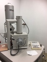
Scanning Electron Microscopy coupled with Energy Dispersive X-ray (SEM-EDS) is a highly useful analytical tool for ceramic studies.
The SEM-EDS technique provides elemental quantification by using X-ray spectroscopy. It offers high-resolution images of ceramic surfaces and provides compositional information on different areas of the same image, which enables analysts to select an area for chemical analysis that is as representative of the original composition as possible. Since energy dispersive X-ray spectrometers provide a quick determination of the elemental composition of a target, compositional elements of ceramic fabrics can be determined by using this technique. Moreover, it provides evidence to evaluate the presence of different stages of vitrification and the temperatures reached during the firing process.
Thanks to these features mentioned above, SEM-EDS can also be used to compare the chemical composition of the body and the surface of the pottery.

SEM schematic.
Source: https://www.britannica.com/technology/scanning-electron-microscope
The scanning electron microscope uses electrons through an electron source to create a grayscale image with good depth of field at high magnification. It has electromagnetic coils referred to as lenses that focus the electron beam to move across a sample and analyse a series of areas.
These electrons move through an electron gun and hit the sample. This interaction creates backscattered electrons, secondary electrons and x-rays, which provide us with the “secondary electron” and “backscattered electron” images.
The secondary electron image (SE) is generally used for the study of microstructure and texture. This includes the identification of the components within the paste such as fine inclusions and clay minerals, while the backscattered electron image (BSE) is used to understand the composition of the materials such as the distribution of areas of different composition within the sample.

EDX schematic.
Source: https://doi.org/10.1016/B978-0-12-809597-3.00446-6
X-rays created through the interaction explained above can be analysed with an Energy Dispersive X-Ray Spectrometer (EDS). Each element within the sample subject produces a series of X-ray peaks in a unique pattern. The EDS technique allows the identification of these peaks and provides a quick determination of the elemental composition of the target hit with the electron gun.
The quality and precision of the analysis depends on a variety of factors, one of which is the sample preparation process. The samples have to be conductive to avoid the build-up of electrons on their surfaces. Therefore, they require special treatment before being put in the high vacuum chamber in the SEM.
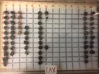
Click HERE to learn about the sample preparation story of the exhibited samples.


